
Blog

Title Image

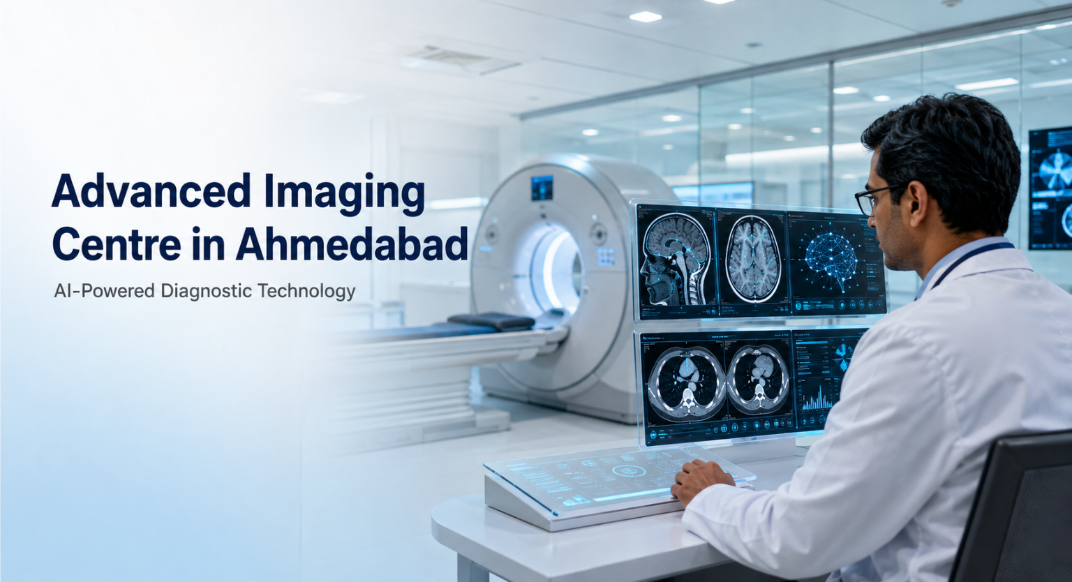
Advanced Imaging Centre in Ahmedabad with AI-powered Technology

Home  /  Blog   /  Advanced Imaging Centre in Ahmedabad with AI-powered Technology
Share At:
Google AI ChatGPT Grok Perplexity

Listen Article

Accurate diagnosis is the foundation of effective medical care. When specialists have access to clear diagnostic images and detailed clinical information, they can investigate medical conditions more precisely and make timely, well-informed decisions about further evaluation or treatment.

Dr. Jivraj Mehta Smarak Health Foundation is proud to introduce its next-generation Advanced Imaging Centre in Ahmedabad, bringing together Artificial Intelligence, deep learning applications and state-of-the-art diagnostic imaging systems.

At the centre of this development is Gujarat’s first Philips Incisive CT 128, equipped with Philips’ advanced AI technologies, including Precise Cardiac and Precise Image. The Advanced Imaging Centre also features Digital X-ray, Wireless Digital Radiography, Philips Affiniti 70 Ultrasound and Full Field Digital Mammography systems.

These advanced diagnostic imaging technologies support superior image clarity, cardiac imaging, breast imaging, elastography, liver assessment, low-dose imaging protocols and intelligent clinical workflows. Together, they empower specialists to support earlier disease detection, improve diagnostic precision and make faster, more confident clinical decisions.

Advanced Diagnostic Imaging in Ahmedabad @ Jivraj Mehta Hospital

Diagnostic imaging helps doctors examine internal organs, tissues, bones and other structures that cannot be evaluated through a physical examination alone. It can play an important role in detecting abnormalities, investigating symptoms, assessing the extent of a condition and monitoring a patient’s response to treatment.

The Advanced Imaging Centre at Dr. Jivraj Mehta Smarak Health Foundation has been developed to provide comprehensive diagnostic imaging supported by modern technology and experienced medical professionals.

By bringing several imaging systems together within one specialised centre, the hospital can support better coordination between radiologists, referring doctors and other specialists. This integrated approach helps clinical teams access relevant diagnostic information and determine the appropriate next step in patient care.

The technologies available at the centre include:

  • Philips Incisive CT 128
  • Precise Cardiac technology
  • Precise Image technology
  • Digital X-ray
  • Wireless Digital Radiography
  • Philips Affiniti 70 Ultrasound
  • Full Field Digital Mammography
  • Elastography and liver assessment capabilities
  • Low-dose imaging protocols
  • AI-enabled intelligent workflow solutions

Each imaging examination is selected according to the patient’s symptoms, medical history and clinical requirements.

Instant WhatsApp Support
WhatsApp Chat Now!

Gujarat’s First Philips Incisive CT 128

A major highlight of the Advanced Imaging Centre is Gujarat’s first Philips Incisive CT 128, equipped with Philips’ AI-enabled technologies.

CT imaging produces detailed cross-sectional images of internal structures within the body. These images provide specialists with important clinical information when investigating different medical conditions.

The Philips Incisive CT 128 combines advanced image acquisition with intelligent processing and reconstruction capabilities. Its AI-supported workflow assists the imaging team at different stages of the examination and helps maintain consistency throughout the scanning process.

The system is designed to support:

  • Detailed diagnostic imaging
  • Advanced cardiac imaging
  • AI-assisted image reconstruction
  • Improved image clarity
  • Cardiac motion correction
  • Efficient imaging workflows
  • Low-dose imaging protocols
  • Faster access to clinically useful images

The Philips Incisive CT platform supports AI-enabled CT Smart Workflow, including technologies developed to improve image reconstruction, cardiac imaging and workflow efficiency.

Advanced Cardiac CT Imaging with Precise Cardiac

Cardiac CT imaging presents a particular challenge because the heart remains in continuous motion. This movement can affect the clarity of the acquired images, especially when detailed visualisation of coronary arteries is required.

Philips Precise Cardiac technology is designed to compensate for cardiac motion during CT imaging. By minimising the effects of movement, the technology can help improve the visualisation of coronary arteries and other important cardiac structures.

Clear cardiac CT images can provide specialists with valuable information when evaluating cardiovascular health. Depending on the patient’s clinical requirements, the imaging findings may help doctors decide whether further evaluation, monitoring or treatment should be considered.

The availability of Precise Cardiac technology strengthens the hospital’s capabilities for advanced cardiac imaging in Ahmedabad. It supports radiologists and cardiac specialists by providing clearer imaging information for clinical interpretation.

Precise Cardiac does not replace the knowledge or judgement of a specialist. It functions as an advanced imaging tool that supports specialists during the diagnostic process.

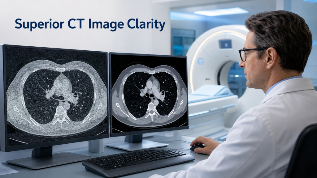
Superior CT Image Clarity with Precise Image

Diagnostic image clarity is essential because even subtle variations may contain clinically important information. Image noise or limited visibility can make interpretation more challenging, particularly when specialists need to examine small structures or low-contrast areas.

Philips Precise Image uses deep learning-based image reconstruction to support high-quality CT imaging. It is designed to reduce image noise and improve the visibility of anatomical structures.

This AI-supported reconstruction technology can help radiologists review CT images with greater clarity and diagnostic confidence. It also supports low-dose imaging protocols, allowing the imaging team to select an appropriate protocol according to the clinical purpose of the examination.

Precise Image contributes to:

  • Clearer reconstructed CT images
  • Reduced image noise
  • Improved visibility of anatomical details
  • Greater consistency in image quality
  • Support for low-dose imaging protocols
  • Efficient reconstruction and image review

By integrating deep learning into CT image reconstruction, the technology supports the balance between image quality, workflow efficiency and responsible dose management.

Low-Dose Imaging Protocols Based on Clinical Requirements

Radiation dose is an important consideration in CT and X-ray imaging. The Advanced Imaging Centre incorporates modern technologies and carefully selected protocols to support patient-centred diagnostic care.

Low-dose imaging protocols are intended to optimise radiation exposure while maintaining the image quality required for clinical interpretation. The appropriate protocol depends on several factors, including:

  • The type of examination
  • The body area being evaluated
  • The clinical purpose of the scan
  • The patient’s medical requirements
  • The image quality needed for interpretation

AI-supported image reconstruction can help the imaging team obtain clinically useful images while following suitable dose protocols.

Low-dose imaging does not mean that every patient or examination receives the same radiation level. Each scan is planned according to its diagnostic purpose and the individual patient’s clinical needs.

Digital X-Ray in Ahmedabad

The Advanced Imaging Centre is equipped with a state-of-the-art Digital X-ray system. Digital X-ray is commonly used to examine bones, joints, the chest and other internal structures based on clinical requirements.

Unlike conventional film-based radiography, Digital X-ray captures images electronically. The acquired images can be processed, stored and made available for clinical review without conventional film development.

Key advantages of Digital X-ray technology include:

  • Rapid image acquisition
  • Clear digital images
  • Efficient image processing
  • Prompt quality assessment
  • Convenient digital storage
  • Faster availability for clinical review

These capabilities can help streamline the diagnostic process while providing doctors with the images required for further evaluation.

Digital X-ray examinations are performed according to the patient’s clinical requirements and interpreted by qualified medical professionals.

Testimonial

“I had a very good and satisfying experience at Jivraj Mehta Hospital. The doctors here are highly experienced and provide clear guidance with utmost care. The hospital environment is clean, well-organized, and efficiently managed by the administrative staff. The nursing team is extremely caring and attentive, ensuring patients feel comfortable throughout their stay. Overall, the patient care is excellent, and I truly appreciate the professionalism and compassion shown by the entire team. Highly recommended!

Flexible Imaging with Wireless Digital Radiography

Wireless Digital Radiography provides greater flexibility during image acquisition. Its wireless detectors can be positioned according to the examination requirements and the patient’s physical condition.

This flexibility can be helpful when conventional positioning is difficult or when a patient has limited mobility. The imaging team can adjust the placement of the detector while maintaining an efficient digital workflow.

Wireless Digital Radiography supports:

  • Flexible detector positioning
  • Efficient image acquisition
  • Prompt availability of images
  • Improved workflow coordination
  • Convenient quality assessment
  • Greater attention to patient comfort

The system complements the centre’s Digital X-ray capabilities and helps imaging professionals adapt the examination process to different clinical situations.

Advanced Ultrasound with Philips Affiniti 70

The Advanced Imaging Centre also features the Philips Affiniti 70 Ultrasound system. Ultrasound uses sound waves rather than ionising radiation to produce real-time images of organs, tissues and other internal structures.

The Philips Affiniti 70 combines advanced image quality with intelligent workflow features. Its real-time imaging capabilities provide specialists with detailed visual information that can support clinical evaluation.

Depending on the patient’s clinical requirements, ultrasound imaging may help specialists:

  • Investigate symptoms
  • Examine organs and soft tissues
  • Assess blood flow
  • Evaluate certain abnormalities
  • Monitor an existing condition
  • Decide whether further investigation is needed

The Philips Affiniti 70 is designed to deliver advanced image quality and efficient workflows across a variety of clinical applications.

Elastography and Advanced Liver Assessment

The Philips Affiniti 70 system supports elastography and liver assessment applications. Elastography is an ultrasound-based technique that helps specialists assess the stiffness of tissue.

Tissue stiffness can provide additional clinical information during the evaluation of the liver and certain other organs. When clinically appropriate, elastography can complement conventional ultrasound findings and support a more comprehensive assessment.

Advanced liver assessment capabilities can help specialists evaluate liver health and determine whether further investigation or monitoring may be needed. Philips identifies automated liver elastography as one of the supported capabilities of the Affiniti 70 platform. Philips Shear Wave Elastography

Elastography findings are not interpreted independently. Specialists consider them together with the patient’s medical history, symptoms, clinical examination and other relevant diagnostic results.

Full Field Digital Mammography in Ahmedabad

The Advanced Imaging Centre is also equipped with a Full Field Digital Mammography system to support advanced breast imaging.

Mammography captures detailed images of breast tissue and can assist specialists in identifying changes that may require further evaluation. Full Field Digital Mammography produces digital images that radiologists can examine closely during screening or diagnostic assessment.

The technology supports:

  • Detailed digital breast imaging
  • Screening based on medical recommendations
  • Evaluation of suspicious breast changes
  • Closer examination of areas of concern
  • Efficient digital image processing
  • Timely clinical review

Digital Mammography may be recommended for routine breast screening or to investigate symptoms such as a breast lump or other unusual changes.

Mammography can support the earlier investigation of breast abnormalities, but it does not guarantee the detection of every condition. A qualified specialist must evaluate the images and may recommend additional examinations when clinically required.

How Artificial Intelligence Supports Radiologists

Artificial Intelligence is increasingly being used in diagnostic imaging to support healthcare professionals during image acquisition, reconstruction and review.

At the Advanced Imaging Centre, AI and deep learning applications complement the experience and clinical judgement of radiologists and imaging specialists.

AI-enabled technologies can assist with:

  • Image reconstruction
  • Image noise reduction
  • Improvement of image clarity
  • Cardiac motion correction
  • Workflow automation
  • Imaging consistency
  • Faster image processing
  • Low-dose protocol support
  • Efficient clinical review

Artificial Intelligence does not diagnose patients independently and does not replace radiologists. The final interpretation remains the responsibility of qualified medical professionals.

Radiologists examine the images and consider them together with the patient’s symptoms, clinical history and other diagnostic information before communicating their findings to the referring doctor.

Patient Benefits of an Advanced Imaging Centre

The next-generation imaging systems at Dr. Jivraj Mehta Smarak Health Foundation are intended to support both diagnostic quality and an efficient patient experience.

Potential patient benefits include:

  • Access to multiple advanced imaging technologies
  • Clear and detailed diagnostic images
  • Improved cardiac imaging capabilities
  • Advanced breast imaging
  • Elastography and liver assessment
  • Low-dose protocols where clinically appropriate
  • Faster imaging workflows
  • Better coordination between specialists
  • Timely availability of images for clinical review
  • More informed clinical decision-making

The precise benefits depend on the examination being performed and the individual patient’s medical condition.

Technology Supported by Clinical Expertise

Advanced imaging equipment delivers its greatest value when supported by trained and experienced healthcare professionals.

At Dr. Jivraj Mehta Smarak Health Foundation, radiologists, imaging technicians and other specialists work together to ensure that examinations are selected and conducted according to each patient’s clinical requirements.

The imaging team focuses on:

  • Following established examination protocols
  • Selecting appropriate imaging techniques
  • Maintaining image quality
  • Supporting patient comfort
  • Reviewing images carefully
  • Communicating relevant findings
  • Coordinating with referring specialists

This combination of advanced imaging technology and professional expertise supports a comprehensive approach to diagnostic care.

Speak with an Expert
Call Call Us Now!

Redefining Diagnostic Excellence at Jivraj Hospital

The next-generation Advanced Imaging Centre at Dr. Jivraj Mehta Smarak Health Foundation reflects our continued commitment to healthcare innovation, diagnostic precision and patient-focused care.

With Gujarat’s first Philips Incisive CT 128 featuring Precise Cardiac and Precise Image, together with Digital X-ray, Wireless Digital Radiography, Philips Affiniti 70 Ultrasound and Full Field Digital Mammography, the centre establishes a new benchmark in cardiac and advanced diagnostic imaging.

Powered by Artificial Intelligence, deep learning applications, superior image clarity, advanced breast imaging, elastography, liver assessment, low-dose imaging protocols and intelligent workflow solutions, these technologies empower our specialists to support earlier disease detection, diagnose with greater precision and make faster, more confident clinical decisions.

Because behind every diagnosis is a life, behind every patient is a family, and behind every timely diagnosis is the promise of a healthier tomorrow.

For more information about available examinations or to schedule an appointment, contact Dr. Jivraj Mehta Smarak Health Foundation in Ahmedabad. The hospital’s existing Radiology Department page provides appointment details and information about its diagnostic services.

Frequently Asked Questions

What facilities are available at the Advanced Imaging Centre in Ahmedabad?

The centre features Philips Incisive CT 128, Digital X-ray, Wireless Digital Radiography, Philips Affiniti 70 Ultrasound and Full Field Digital Mammography. It also supports cardiac imaging, elastography, liver assessment and low-dose imaging protocols.

What is Philips Incisive CT 128?

Philips Incisive CT 128 is an advanced CT imaging system equipped with AI-supported technologies. At Dr. Jivraj Mehta Smarak Health Foundation, it includes Precise Cardiac and Precise Image technologies for advanced cardiac imaging and deep learning-based image reconstruction.

How does Precise Cardiac support cardiac CT imaging?

Precise Cardiac is designed to compensate for cardiac motion during CT imaging. This can improve the visualisation of coronary arteries and other cardiac structures, supporting more confident clinical interpretation.

What is Precise Image technology?

Precise Image is a deep learning-based CT image reconstruction technology. It is designed to reduce image noise, improve anatomical visibility and support high-quality imaging with suitable low-dose protocols.

Does the Advanced Imaging Centre offer Digital X-ray?

Yes. The centre has Digital X-ray and Wireless Digital Radiography systems that support efficient image acquisition, digital processing, convenient storage and prompt clinical review.

What is the benefit of Wireless Digital Radiography?

Wireless Digital Radiography offers flexible detector positioning. This can make image acquisition more adaptable for patients with limited mobility or examinations requiring alternative positioning.

Is advanced ultrasound available at Jivraj Hospital?

Yes. The Advanced Imaging Centre features the Philips Affiniti 70 Ultrasound system, which supports advanced image quality, real-time imaging and intelligent clinical workflows.

What is elastography?

Elastography is an ultrasound-based technique used to assess tissue stiffness. It can provide additional clinical information during liver assessment and the evaluation of certain other organs when medically appropriate.

Is Digital Mammography available at the centre?

Yes. Full Field Digital Mammography is available to support detailed digital breast imaging, screening and the evaluation of suspicious breast changes.

Does Artificial Intelligence replace the radiologist?

No. AI supports image reconstruction, motion correction, image clarity and workflow efficiency. Qualified radiologists remain responsible for interpreting the images and reporting the findings.

Are low-dose imaging protocols available?

Yes. The centre supports low-dose imaging protocols where clinically appropriate. The selected protocol depends on the examination type, diagnostic purpose and individual patient requirements.

How can patients book a diagnostic imaging appointment?

Patients can contact Dr. Jivraj Mehta Smarak Health Foundation or use the appointment facility available through the hospital’s website to enquire about diagnostic imaging examinations.

<< 1 >>